When it comes to understanding how your brain communicates with your body, neurologists have a variety of tools at their disposal. One of the most precise and informative is Evoked Potential (EP) testing, a non-invasive procedure that measures how your nervous system responds to specific stimuli. By evaluating the function of nerve pathways, EP testing can reveal subtle problems even before symptoms appear, making it a valuable diagnostic tool for conditions like multiple sclerosis, optic neuritis, or nerve pathway disorders. Unlike MRI scans, which show structure, EP testing shows function, giving neurologists a clearer picture of your nervous system health.
Continue reading and find out how non-invasive, painless EP tests can uncover subtle nerve or brain pathway problems.
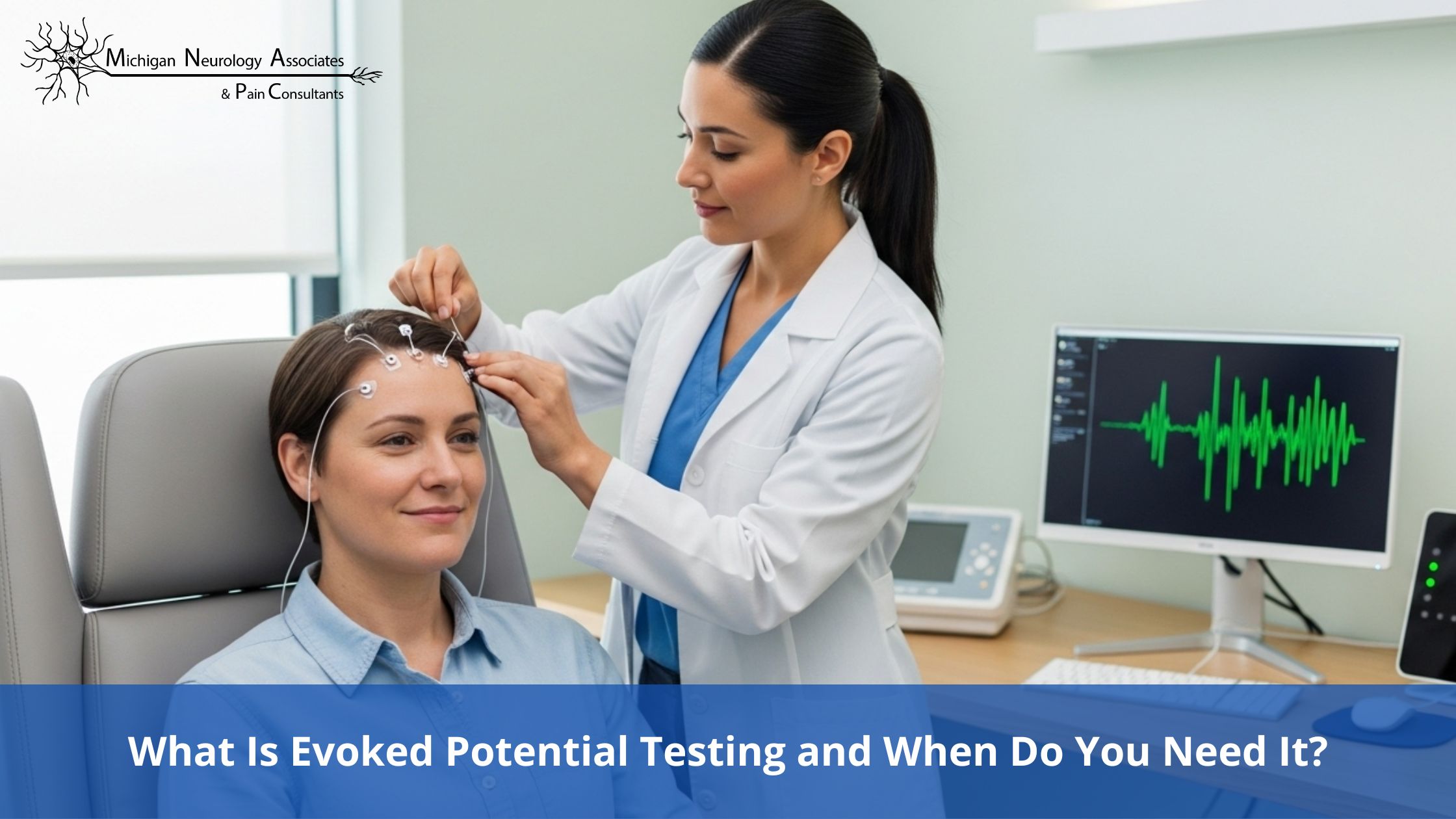
Evoked potential testing measures the electrical activity of your brain in response to stimuli such as light, sound, or touch. During the test, small electrodes are placed on your scalp or skin, and your brain’s response to these signals is recorded.
The process is painless, quick, and non-invasive, typically lasting between 30 to 60 minutes. While an MRI provides a detailed image of the brain or spinal cord structure, EP testing reveals how well these structures function. This functional insight can be critical in diagnosing neurological conditions that may not yet produce visible changes on imaging.
There are several types of EP tests, each designed to evaluate different sensory pathways:
Neurologists choose the type of EP test based on the patient’s symptoms and suspected condition.
EP testing is particularly useful in situations where other tests may be inconclusive or early detection is critical. Common scenarios include:
EP testing is often ordered when MRI results do not provide a complete picture, making it an essential tool in the neurologist’s diagnostic toolkit.
Preparing for an EP test is simple. Patients should:
It is important to remember that the test is safe, non-invasive, and does not require anesthesia or recovery time.
During the test, you can expect:
The procedure is entirely painless and is conducted in an outpatient setting with no recovery time needed.
Normal EP responses indicate healthy nerve pathways, while abnormal responses may suggest delays or blockages in nerve signal transmission. These findings can help neurologists:
Neurologists will review the results with you in clear, understandable terms, ensuring you know exactly what the findings mean for your health.
EP testing offers several advantages:
By identifying problems early, EP testing can help prevent complications and support timely treatment.
Evoked potential testing is a powerful, non-invasive tool that helps neurologists uncover hidden issues in your brain and nerve pathways, often before symptoms appear. By providing a clear picture of how your nervous system functions, it complements other diagnostic tools like MRI and guides timely treatment. If you’re experiencing unexplained vision, hearing, or nerve symptoms, consulting a neurologist about EP testing could be a crucial step toward better neurological health.
When it comes to your nervous system, knowledge is power. At Michigan Neurology Associates & Pain Consultants, we offer precise, painless evoked potential testing to assess how your nerves and brain are functioning. With expert guidance and personalized care, you can take informed steps toward early detection and treatment. Schedule your consultation today and take charge of your health.